Which eye movement has the longest latency and fastest velocity respectively?
Accommodation / Vergence / Oculomotor Function
Optics
No
U
D
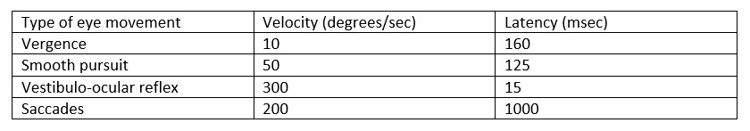
Please refer to the table below that describes the latencies and
velocities of the various eye movements. From this table, you can see
that saccades have both the longest latency (200 msec) and fastest velocity (1000 degrees/sec).
Vergence
movements align the eyes to maintain bifoveal fixation of an object so
that fusion is obtained. Vergence movements involve BOTH eyes (in
contrast to "ductions" which move only one eye). Convergence is the
movement that occurs when both eyes rotate inwards. This is stimulated
by relative movement that brings an object closer to the observer. In
contrast, divergence is the movement that occurs when both eyes rotate
outwards. This is stimulated by relative movement that brings an object
farther from the observer.
The smooth pursuit and saccadic systems
are controlled by different anatomical pathways that converge at the
level of the brainstem. The purpose of the smooth pursuit system is to
hold a target image steady on the fovea during linear motion of the
object. The purpose of the saccadic system is to rapidly bring the
object of interest to the fovea.
The vestibulo-ocular reflex (VOR) holds the retinal image of an object steady during brief head
rotation or translation. It achieves this by producing eye movements
in a direction opposite to those of head acceleration. However, during prolonged head rotation/translation (e.g. following a moving object for a long time), the VOR weakens quickly and is supplemented by the optokinetic nystagmus (OKN) system.
The OKN system uses smooth pursuit to track a moving object, but then
introduces a saccade in the opposite direction if the object is moving
too fast for the pursuit or if the maximum amplitude of the pursuit is
reached.
Heads up! You can use keyboard for test navigation: press → for Next,
← for Previous, M for Mark/Unmark, P for Pause,
R for Review, A,B,C,... or 1,2,3,... to select answer.